2021年10月18日
骨科植入物手术包括人造关节置换,脊柱仪器和断裂修复。尽管这种程序可以改善生活质量,但它们有并发症的风险。一种纸Sasamoto,T等人。对此进行扩展;本文仔细研究了该论文。
一个严重的问题是术后植入物感染。事件速率是显着的,并且正在研究培养金,银,碘或抗生素(如庆大霉素或万古霉素)到植入物的金属表面的各种技术,目的是预防植入物周围感染。
Diamond-like carbon (DLC) is a reference term for carbon thin films possessing high hardness, electrical insulation, and infrared transmission. The combination of F-DLC with fluorine has an anticoagulant effect, finding application in clinical settings and blood vessel stents. The researchers reported excellent antibacterial properties expressed by coating.
关注的一个重要原因是对DLC涂层引起的成骨的影响。研究人员研究了F-DLC涂层对成骨的影响,同时与未涂层的钛合金进行比较。
This article measures the release of F-ions from the F-DLC coating. F-ions can increase both direct cytotoxicity and metabolic toxicity and are also capable of bonding with calcium and magnesium in the body to cause hypocalcemia or hypomagnesemia.
Methodology
The study investigated the effects of F-DLC coated titanium alloy in vivo osteogenesis with the approval of the Animal Research Ethics Review Board. The experiment focused on local bone response rather than the system response.
The test specimens were of Ti-6Al-4V metal alloy, comprising 90% titanium, 6% aluminum, 4% vanadium by a mass fraction. The high bio-affinity of the alloy enables broad application potential in bone-anchoring implants.
射频电感耦合等离子体化学蒸气沉积(CVD)技术用于铺设三层涂层。各个层是碳化硅(SIC),DLC涂层(A-C:H结构)和F-DLC涂层(23 at。%F+A-C:H:H:F结构)。
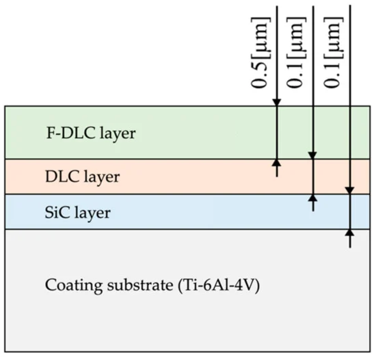
图1。涂层厚度和结构。图片来源:Sasamoto等,2021
将测试片形成直径为5 mm的固体柱,长度为20毫米。平行缝隙宽0.5毫米,每列沿着12个位置的短轴以1毫米的间隔在短轴上进行2 mm的深处。
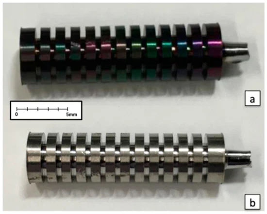
图2。(a)覆盖有氟钻石样碳(F-DLC)的植入物;(b)无F-DLC涂层的控制植入物。图片来源:Sasamoto等,2021
一种fter administering anesthesia to dogs, following other standard procedures, a 10-cm skin incision was made in the anterior lateral margin of the femur. The initial cut was made between the quadriceps femoris muscle and tensor fascia lata muscle.
通过将前向牵引力放在股股骨肌肉肌肉上,暴露于肌椎管。使用ϕ5.0 mm钻头在股骨的diaphysy区域(diaphessy的中心,靠近中心2 cm)和中心远端2 cm的肌椎管区域的长轴沿3个位置创建孔。
The diameter of each hole was then enlarged using a ϕ5.1 mm reamer. During this process, the drill and reamer were cooled continuously to minimize irritation to the bone. More saline solution was used to remove the residual tissue. A total of six implants from Group F or Group C were inserted.
一种fter 4, 8, and 12 weeks of the implantation, the animals were sacrificed by intravenous injection of KCL, and the specimens were collected.
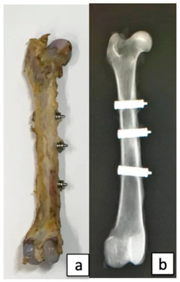
图3。((a) Collected femur; (b) Radiograph of the femoral bone. Image Credit: Sasamoto et al., 2021
一种t every interval, eight femurs were collected. Double staining was done and calcein was injected subcutaneously before collecting the bone specimens. Under fluorescence, bones calcified before and after implantation were differentiated.
然后,用70%乙醇固定骨头,切成三块。将所有碎片均抛光并用Villanueeva骨色骨染色染色,以形成横截面股骨标本。每个收集周总共创建了72个标本,并使用24个标本。
自然光,偏振光和荧光光用于检查最大程度影响骨骼的皮质骨。荧光光用于评估新形成的组织的成熟度。
Bone histomorphometry was then applied to measure eight parameters, namely, bone mass (%), trabecular thickness (µm), osteoclast number (N/mm), resorption area (%), osteoblast number (N/mm), osteoid area (%), calcification rate (µm/day), the annual rate of bone formation (µm/year).
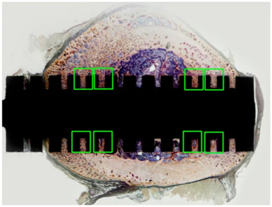
图4。抛光的非矿化股骨标本(整个标本)的横截面。病理检查和骨骼组态计量学评估集中在绿色盒子区域,在该区域,皮质骨接触植入物的缝隙部分,因为预计骨形成在这些区域会更加活跃。图片来源:Sasamoto等,2021
The F-ion elution was confirmed using solid round columns of 25 mm diameter and 5 mm thickness and polished to a mirror finish (Ra = 0.1 µm). Testing was performed by lanthanum–alizarin complex one absorption spectrometry complying methods.
相应地施加了涂层,并在模仿体内条件下,浸没在防御紫外线的暗室中进行。设置了37°C±0.2°C的温度以保持理想状态。将测试片留在恒温室内的干燥器中的普通盐水溶液中一定时间。
浸泡溶液后来用作测试溶液,浸泡了2周,4周和8周,并在三个实例上进行了测试。
The student’s t-test was used for statistical analysis, to compare Group F and Group C for F ions elution. All data were shown as mean values for each group ± standard deviation. The significance level was set at p < 0.05. Results were graphed with standard deviations.
Results
图5描绘了自然光线下的观察结果。在第4周,确认两组纤维骨组织的形成。在第8周,在C组中可见成纤维细胞和纤维骨素,而在F组中,小梁菌在狭缝中形成深处,并且看不到未成熟的细胞。在第12周,在C组的缝隙中有深处的小梁形成,但注意到没有未成熟的细胞。小梁在F组中比在C组中更密集。
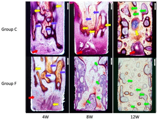
图5。自然光线下缝隙内表面的微观图像。黄色箭头表示纤维骨质骨,蓝色箭头是纤维骨头,红色箭头是成纤维细胞,绿色黄色箭头表示层状骨骼。图片来源:Sasamoto等,2021
图6显示了在偏光光下对样品的观察,其中层状骨清晰可见。在第8周,层状骨和骨骨被可见,但在第12组中看不到。在第12周,比C的层状骨和骨比。
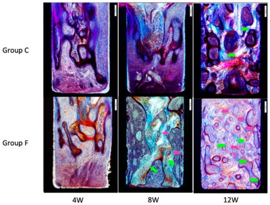
图6。偏光光的缝隙内表面的微观图像。绿色黄色箭头表示层状骨,粉红色箭头表示骨。在第8周的F组中可见层状骨和骨。
In Figure 7, the observations under the fluorescent light of the specimen are presented. Newly formed bone tissue with calcein green was identified. In both F and C groups, the calcein-labeled areas increased from week 8 to week 12. At Week 12, double labeling of specimens was noted in Group F.
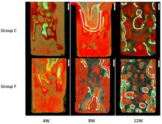
图7。荧光灯下缝隙内表面的微观图像。新形成的骨组织用钙软蛋白绿色标记鉴定出来。白色箭头显示双重标签的区域。在第12周,在F组中形成的骨头比组的骨骼多。图片来源:Sasamoto等,2021
骨组织形态计量学的结果如图8A – H和表1所示。F组F中的平均骨体积/组织体积/组织体积±SD明显增加。小梁F的平均厚度±SD显着增加。骨表面±SD的降低明显更大。
平均侵蚀的表面/骨表面±SD的F组降低明显更大。在8周时,F组的骨形成速率/组织体积的比率明显更大。
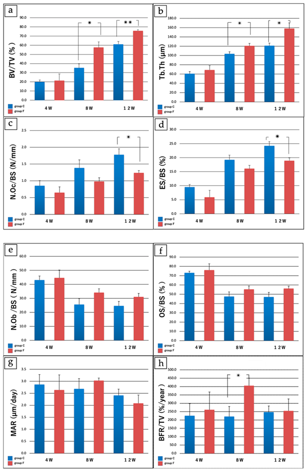
Figure 8.骨骼组态法的结果。(a)骨体积/组织体积(%)。在第4周,两组之间没有明显差异。在第8周,C组的骨体积/组织体积(%)为35.37±8.70,F组为57.53±12.4(P <0.05),第12周的C组为61.17±2.12,组为75.77±5.88。F(p <0.005),F组F。(b)小梁厚度(µm)的增加明显更大。在第4周,两组之间没有明显差异。在第8周,小梁的厚度为103.71±8.8 µm,在F组中为120.87±10.5 µm(P <0.05),第12周,组为121.19±11.1 µm,C组为158.11±24.2 µm,组为158.11±24.2 µm。F(p <0.05),F组F。(c)破骨细胞/骨表面长度(N/mm)的数量明显更大。在第4周和第8周,两组之间没有明显差异。组C组的破骨细胞/骨表面长度为1.78±0.35/mm,F组为1.28±0.13/mm(P <0.05)(P <0.05),F组f。 (%). At Week 4 and Week 8, no significant differences were noted between groups. At Week 12, eroded surface/bone surface (%) was 24.19 ± 3.03 in Group C vs. 18.94 ± 1.98 in Group F (p < 0.05), with a significantly greater decrease in Group F. (e) Number of osteoblasts/bone surface length (N/mm); (f) Osteoid surface/bone surface (%). (g) Mineral apposition rate (µm/day). (h) Bone formation rate/tissue volume (%/year). For (e–g), no significant differences were found between Group C and Group F at any time point. For (h), the ratio of bone formation rate/tissue volume at 8 weeks was 220.1 ± 119.08 %/year in Group C vs. 405.47 ± 92.29 %/year in Group F (p < 0.05), with a significantly greater increase in Group F. * p < 0.05; **p < 0.005 for (a–d,h). Image Credit: Sasamoto et al., 2021
表格1。骨骼组态法的结果。Source: Sasamotoet al。,2021年
|
4W (Groups
C vs. F) |
8 W (Groups
C vs. F) |
12 W(组
C vs. F) |
| 答:骨体积/组织体积(%) |
19.89±4.51 vs. 21.51±14.1 |
35.37±8.70 vs。
57.53 ± 12.4 * |
61.17±2.12 vs. 75.77±5.88 ** |
| B:小梁厚度(μm) |
60.71 ± 9.10 vs. 68.92 ± 18.8 |
103.71 ± 8.8 vs. 120.87 ± 10.5 * |
121。19 ± 11.1
158.11±24.2 * |
| C:否则破骨细胞/骨表面(N/mm) |
0.85 ± 0.30 vs. 0.65 ± 0.34 |
1.39±0.48 vs。
0.98±0.22 |
1.78±0.35 vs. 1.28±0.13 * |
| D:侵蚀的表面/骨表面(%) |
9.47±1.87 vs. 5.88±4.89 |
19.27±3.23与16.08±2.36 |
24.19±3.03 vs. 18.94±1.98 * |
| e: No. osteoblasts/Bone surface (N/mm) |
43.10±5.78 vs. 44.68±11.11 |
25.62±8.51 vs. 34.15±5.34 |
24.66±6.35 vs。
31.08±4.90 |
| F:骨骼表面/骨表面(%) |
73.10±3.40 vs. 76.00±13.50 |
47.60±9.84 vs. 55.31±7.14 |
47.10±9.90 vs. 56.17±5.14 |
| G:矿物质申请率(/天μm) |
4.87±0.83 vs. 2.64±1.25 |
2.69±0.86 vs。
3.03±0.21 |
2。41± 0.52 vs. 2.08 ± 0.69 |
| H:骨形成率/骨头体积(%/年) |
225。50 ± 145.50 vs. 261.67 ± 211.94 |
220.91±119.08 vs. 405.47±92.29 * |
247.09±73.64与254.80±142.83 |
* p <0.05;** p <0.005。
图9显示了沉浸周期(几周)的测试片和氟离子洗脱体积的值。氟离子的数量随时间增加。
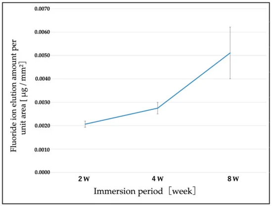
Figure 9.随着时间的流逝,每单位区域的氟洗脱量随时间变化。F离子的浓度随着时间而增加。图片来源:Sasamoto等,2021
讨论
The study established a comparison between traditional titanium alloy implants and F-DLC-coated titanium alloy implants to determine the effects of F-DLC coating on bone formation in vivo. The findings show the bone union was more accelerated with F-DLC coated implants than with non-coated titanium alloy implants.
还可以观察到骨骼联合通过没有氟的DLC涂层大大促进了骨结合。此外,补充实验肯定了F-DLC涂层随着时间的流逝。在第12周,由于F离子的溶液较少,破骨细胞会减少。
Excessive or prolonged inflammation was related to extended cell and callus proliferation. As per the study, the selected F ions formed sodium fluoride leading to anti-inflammatory action that shortened the duration of inflammation during the early bone union.
植入物仅测试8周。该研究是对Beadle Dogs进行的,但还需要人类研究。即使有这些局限性,该研究也肯定了F-DLC涂层的抗菌特性。
结论
该研究揭示了通过F-DLC涂层合金促进骨骼生长的促进,并且合金在比报道的氟化物毒性水平低的水平上洗脱。考虑到抗菌和骨骼联合促进特征,F-DLC表现出有望作为骨植入物的涂料材料。
期刊参考:
Sasamoto, T., Kawaguchi M., Yonezawa K., Ichiseki T., Kaneuji A., Shintani K., Yoshida A., Kawahara N. (2021) Antibacterial Fluorinated Diamond-Like Carbon Coating Promotes Osteogenesis—Comparison with Titanium Alloy.应用科学欧洲杯线上买球。可用网址:doi.org/10.3390/App11209451。
References and Further Reading
Davne, S H & Myers, D L (1992) Complications of lumbar spinal fusion with transpedicular instrumentation.脊柱, 17 (Suppl. 6), pp. S184–S189.doi.org/10.1097/00007632-199206001-00021。
Sponseller, P. D.,et al。(2000)神经肌肉脊柱侧弯手术后的深层伤口感染:危险因素和治疗结果的多中心研究。脊柱,25,第2461–2466页。doi.org/10.1097/00007632-200010010-00007
Sperling,J。W.,et al。(2001)肩关节置换术后感染。临床骨科和相关研究, 382, pp. 206–216.doi.org/10.1097/00003086-200101000-00028。
Minnema,B。et al。(2004)主要全膝关节置换术后手术部位感染的危险因素。感染控制与医院流行病学,25,第477–480页。doi.org/10.1086/502425。
Bengtson, S & Knutson, K.等。(1991)被感染的膝关节置换术。357例案件的6年随访。一种cta Orthopaedica Scandinavica,62,第301–311页。doi.org/10.3109/17453679108994458。
Hill,G e&Droller,D G(1989)使用抗菌预防剂未元素的总髋关节置换后的急性和亚急性深感染。Orthopedic Reviews, 18, pp. 617–623.doi.org/10.3390/antibiotics9080495。
Wymenga,A。B.,et al。(1992)与关节置换术后脓毒症关节炎相关的围手术因素。362膝盖和2,651个髋关节手术的预期多中心研究一种cta Orthopaedica Scandinavica, 63, pp. 665–671.doi.org/10.1080/17453679209169732。
Phillips,C。B.,et al。(2003)在选择性总髋关节置换后的前六个月,位错率,肺栓塞和深层感染的发生率。骨与关节手术杂志,85,第20-26页。doi.org/10.2106/00004623-200301000-00004。
卢克,M。et al。(2003)金属植入物的庆大霉素涂层减少了大鼠与植入物相关的骨髓炎。Bone,32,第521–531页。doi.org/10.1016/S8756-328200050-4。
Antoci,V。,et al。((2007) Vancomycin bound to Ti rods reduces periprosthetic infection: Preliminary study.临床骨科和相关研究, 461, pp. 88–95,doi.org/10.1097/blo.0b013e318073c2b2。
Neut, D. A.,et al。(2015)可生物降解的庆大霉素 - 羟基磷灰石涂层可预防无胶结髋关节假体。欧洲细胞和材料欧洲杯足球竞彩,2,第42-55页,doi.org/10.22203/ecm.v029a04。
Noda, I.,et al。(2009)新型的热喷涂抗菌涂层和银离子释放性能的评估。Journal of Biomedical Materials Research, Part B, 89B, pp. 456–465,doi.org/10.1002/jbm.b.31235。
Funao, H.,et al。((2016) A novel hydroxyapatite film coated with ionic silver via inositol hexaphosphate chelation prevents implant-associated infection.Scientific Reports,6,p。23238,doi.org/10.1038/SREP23238。
Ando,Y。,et al。((2010) Calcium phosphate coating containing silver shows high antibacterial activity and low cytotoxicity and inhibits bacterial adhesion.欧洲杯足球竞彩材料科学与工程c欧洲杯线上买球,30,第175-180页,doi.org/10.1016/j.msec.2009.09.015。
Zainali,K.,et al。(2009)金涂层对实验植入物固定的影响。Journal of Biomedical Materials Research。A,88,第274-280页,doi.org/10.1002/jbm.a.31924。
Shirai,T.,等。(2011)抗菌碘支持的钛植入物。Acta Biomater,第7页,第1928- 1933年,doi.org/10.1016/j.actbio.2010.11.036。
Stuart,B。W.,et al。((2021) New solutions for combatting implant bacterial infection based on silver nano-dispersed and gallium incorporated phosphate bioactive glass sputtered films: A preliminary study.Bioactive Materials, 8, pp. 325–340,doi.org/10.1016/j.bioactmat.2021.05.055。
Bee,S.-L。,et al。(2021)与生物活性和抗菌特性的银纳米颗粒二十二磷灰石纳米复合材料合成。材料科学杂志:医学材料欧洲杯足球竞彩欧洲杯线上买球,32,第106页,doi.org/10.1007/s10856-021-06590-y。
Aisenberg,S&Chabot,R(1971)钻石状碳薄膜的离子束沉积。材料科学杂志:医学材料欧洲杯足球竞彩欧洲杯线上买球, 42, p. 2953,doi.org/10.1063/1.1660654。
T. Saito,et al。(2005)氟化钻石样碳膜的抗巩固性。钻石和相关材料欧洲杯足球竞彩,14,第1116–1119页,doi.org/10.1016/j.diamond.2004.09.017。
Yonezawa,K。,et al。((2020) Evaluation of antibacterial and cytotoxic properties of a fluorinated diamond-like carbon coating for the development of antibacterial medical implants.一种ntibiotics, 9, p. 495.doi.org/10.3390%2Fantibiotics9080495。
我的,Y。,et al。(2014)仿生钻石样碳涂层钛对体外成骨细胞和破骨细胞分化的影响。光聚合剂科学与技术杂志欧洲杯线上买球。2014,27,第373–378页。doi.org/10.2494/photopolymer.27.373。
我的,Y。et al。(2012)仿生钻石样碳涂层钛在体外抑制RANKL依赖性破骨细胞分化。光聚合剂科学与技术杂志欧洲杯线上买球,25,第523-528页。doi.org/10.2494/photopolymer.25.523。
Shuto, T.,et al。(2016)在氢化四面体非晶碳涂层钛上的成骨细胞和破骨细胞的分化。光聚合剂科学与技术杂志欧洲杯线上买球,第29页,第413–418页。https://doi.org/10.2494/photopolymer.29.413。
日本工业标准:JIS K 0102:2016。工业废水的测试方法;一般公司协会工业环境管理协会;日本标准协会:日本东京,2016年;第108–114页。
Kawaguchi,M。,et al。((2021) Bone formation at Ti-6Al-7Nb scaffolds consisting of 3D honeycomb frame and diamond-like carbon coating implanted into the femur of beagles.Journal of Biomedical Materials Research,109,第1283–1291页,doi.org/10.1002/jbm.b.34789。
Farley,J。R.,et al。((1983) Fluoride directly stimulates proliferation and alkaline phosphatase activity of boneforming cells.欧洲杯线上买球,222,第330–332页。doi.org/10.1126/欧洲杯线上买球science.6623079。
玛丽,P。J。,et al。(1992)刺激骨质疏松症患者的骨形成,该患者在体外通过成骨细胞通过成骨细胞的DNA合成增加而刺激了骨形成。骨与矿物研究杂志, 7, pp. 103–113.doi.org/10.1002/jbmr.5650070115。
Khokher,M A&Dandona,P(1990)氟化物刺激人类成骨细胞刺激[3H]胸苷掺入和碱性磷酸酶。Metabolism,39,第1118–1121页。doi.org/10.1016/0026-0495(90)90081-m。
Wergedal, J. E.,et al。(1988)氟化物和牛骨提取物影响人骨细胞培养物中的细胞增殖和磷酸酶活性。临床骨科和相关研究,233,第274–282页。doi.org/10.1097/00003086-198808000-00034。
Taves, D R (1970) New approach to the treatment of bone disease with fluoride.联邦诉讼, 29, pp. 1185–1187.
Messer, H. H.,et al。((1973). Fluoride, parathyroid hormone and calcitonin: Effects on metabolic processes involved in bone resorption.Calcified Tissue Research,13,第227–233页。doi.org/10.1007/bf02015412。
McCann,H。G.,et al。(1953)氟离子与羟基磷灰石的反应。生物化学杂志, 201, pp. 247–259.
刘,S。et al。(2019)含氟的羟基磷灰石通过抑制破骨细胞的分化和体外和体内功能来抑制骨吸收。细胞增殖,52,p。E12613。doi.org/10.1111/cpr.12613。
Rich, C & Feist, E (1970) The Action of Fluoride on Bone. InFluoride in Medicine; Vischer, T., Ed.;Hans Huber出版物。瑞士伯尔尼,第70-87页。doi.org/10.3390/App11209451。
Hedlund,L&Gallagher,JC(1989)用氟化钠治疗的骨质疏松妇女的髋部骨折发生率增加。骨与矿物研究杂志,4,第223–225页。doi.org/10.1002/jbmr.5650040214。
Varughese,K&Moreno,E C(1981)含有氟化物的稀溶液中钙磷灰岩的晶体生长。钙化组织国际, 33, pp. 431–439.doi.org/10.1007/bf02409467。
Eanes, E D & Reddi, A H (1979) The effect of fluoride on bone mineral apatite.Metabolic Bone Disease and Related Research,第2页,第3-10页。
Moreno, E. C.,et al。(1977)与龋齿研究相关的氟化物 - 磷灰石系统的物理化学方面。Caries Research,11,第142-171页。doi.org/10.1159/000260299。
Bhawal,K。U.,et al。(2020)用低水平氟化钠处理伤口愈合和骨髓间充质干细胞的成骨分化。Dental Traumatology, 36, pp. 278–284,doi:10.1111/edt.12532。
Brown, J. H.,et al。(1972)氟化钠的抗炎活性。Archives Internationals de Pharmacodynamie et de Therapie, 195, pp. 361–371.
Lee, H J & Choi, C H (2015) Anti-inflammatory effects of bamboo salt and sodium fluoride in human gingival fibroblasts—An in vitro study. Kaohsiung.国际医学杂志欧洲杯线上买球, 31, pp. 303–308.doi.org/10.1016/j.kjms.2015.03.005。
医学研究所(美国)饮食参考摄入科学评估的常务委员会(1997)钙,磷,镁,维生素D和氟化物的饮食参考摄入量;国家科学院出版社:华盛顿特区,美国;第288–313页。doi.org/10.17226/5776。