冷冻电子显微镜(Cryo-EM)越来越被用作主流技术,以研究分子分辨率下的蛋白质组件,病毒和细胞的结构。本文重点介绍了冷冻EM的不同方面,包括其优势和缺点,应用,冷冻EM和电子显微镜(EM)技术之间的差异以及涉及冷冻EM技术的最新研究。
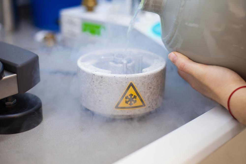
Scientist preparing samples for Cryo-electron microscopy in liquid ethane under liquid nitrogen temperature. Image Credit: PolakPhoto/Shutterstock.com
什么是冷冻EM?
冷冻EM被称为EM的一种形式,其中使用透射电子显微镜(TEM)在低温条件下获得对辐射敏感的标本的图像。短语冷冻EM通常用于几种实验方法,例如单粒子冷冻EM,电子晶体学和冷冻电子层析成像。
每种冷冻EM方法都可以单独使用,并在混合方法中使用,其中从冷冻EM获得的信息与从核磁共振(NMR)光谱和X射线晶体学方法获得的互补信息合并。当前,冷冻EMS变得更容易获得,并且更易于使用,并用于成像生物标本,例如细菌,暴发细胞和完整的组织切片。
EM和Cryo-EM之间的差异
Conventional EM methods use staining, dehydration, and chemical fixation of specimens to obtain their image. The interaction between the organic matter and electrons extensively damages the biological specimens in EM.
Meanwhile, cryo-EM preserves the native hydrated state of specimens, as this technology does not require additional methods for imaging specimens.
The electron irradiation breaks the chemical bonds within the specimens, which leads to the generation of free radicals that further cause secondary damage. Although low electron doses can preserve specimens by minimizing the radiation damage, such low doses lead to noisy images.
冷冻EM可以有效地解决此问题,因为该技术的成像是使用在液态氦或液氮温度下保持的冷冻样品进行的,从而降低了电子辐照对样品的不利影响。
生物标本在玻璃样冰层中迅速玻璃化,然后在液氦气和/或液氮温度下成像。在氮温度下,辐射损伤大大降低,这允许使用较高的电子剂量来获得具有良好信噪比的图像。液氮和氦气也可用于通过冷冻EM方法在接近分子水平的样品中获得三维(3D)的重建。
冷冻EM的优势和缺点
冷冻EM技术不需要晶体,适用于成像蛋白及其大分子量复合物以及膜蛋白的结构分析。此外,该技术还保留了生物样品的功能状态和天然活性,并解决了其他技术(例如X射线晶体学)未解决的结构。
但是,冷冻EM对于成像非常小的蛋白质无效,需要大量时间来生成样品图像。此外,该技术需要非常高的样品均匀性,这增加了获得柔性蛋白的高分辨率图像的困难。此外,当前的冷冻EM分辨率不足以用于药物研究和开发,因为该技术在某些情况下通过该技术获得的图像具有较低的信噪比。
冷冻EM方法的主要应用
Cryo-electron tomography is used to create 3D structures of macromolecular assemblies based on a series of images, with every image obtained at a different tilt relative to the incident electron beam direction. However, imaging of thicker specimens, such as eukaryotic cells or large viruses, is often performed using an imaging filter that enhances the contrast by removing inelastically scattered electrons.
然后将这些图像通过计算结合以生成断层图,这主要是样品的3D图像。从这些层析扫描研究中出现了几种见解,例如膜蛋白组件的横向排列和细菌细胞骨架的组织。冷冻电子断层扫描的其他应用包括研究不同的细胞骨架和丝状组件以及薄哺乳动物细胞区域的成像。
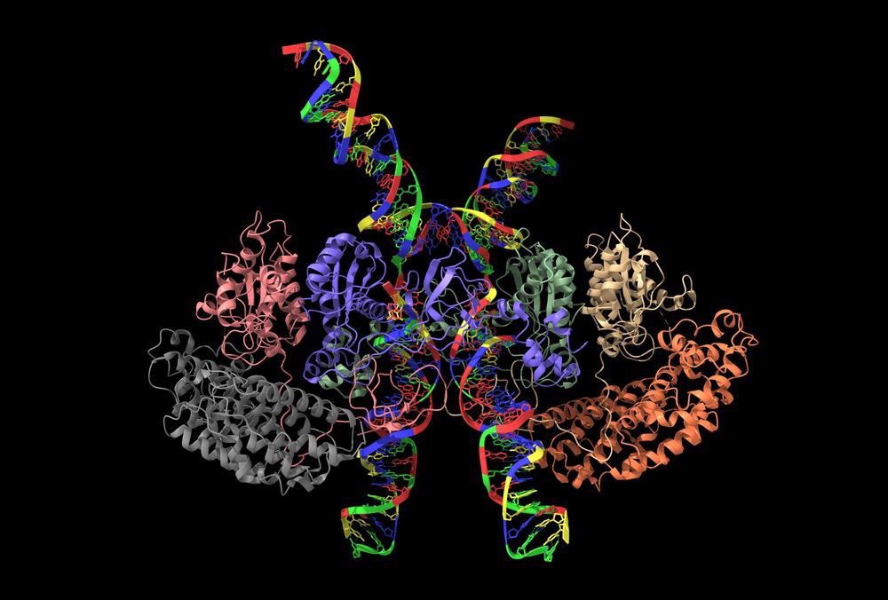
人类T细胞白血病病毒类型-1(HTLV-1)内体的冷冻EM结构。图片来源:Volodymyr dvornyk/shutterstock.com
单粒子分析是最常用的冷冻EM技术。在单粒子分析中,从几个二维(2D)投影图像中获得的数据,这些图像具有各种取向中蛋白质复合物的匹配副本,以产生生物结构的3D重建。在成像二十面体病毒时实现了使用单粒子方法的最高分辨率。单粒子成像的其他应用包括在10个动态蛋白质复合物的10个埃斯特罗姆分辨率下重建以及构象异质样本的分析。
Recent Studies Using Cryo-EM
最近,Cryo-EMS广泛用于收集蛋白质和其他分子结构的图像,并将其存储在电子显微镜数据库(EMDB)中,这是一种流行的EM分解结构的存储库。预计这些结构将用于鉴定蛋白质的工作及其在疾病诊断和治疗中的作用。
Cryo-EM was also used to identify the structural changes in coronavirus disease 2019 (COVID-19) variants to understand the evolution of the virus, which increased their infectivity. High-resolution cryo-EM was employed to detect the changes in the spike protein structure of beta and alpha variants. The research was published in the journal自然通讯。
此外,使用冷冻EM首次识别细胞过程的复杂功能。研究人员,发表在《杂志》上细胞发现,使用冷冻EM密切观察脑细胞中存在的ATP13A2蛋白。该蛋白质导致神经发生疾病,例如帕金森氏病,因为它因遗传突变而失去功能。在成像过程中,将样品冷冻至-238°Fahrenheit和-460°Fahrenheit之间的温度。
结论
To summarize, cryo-EM has emerged as one of the effective imaging techniques to determine the structures of biological assemblies and macromolecules. In the future, cryo-EM can be potentially used for structure-based drug discovery. However, more research is required to use the technique for imaging membrane protein complexes and low-copy-number macromolecules in their native environment.
来自AZOM的更多信息:电子光谱仪中的微通道板检测器
References and Further Reading
Subramaniam,S.,Patwardhan,A.,Pierson,J。et al。冷冻电子显微镜:非微观镜家的底漆。FEBS期刊2013。https://febs.onlinelibrary.wiley.com/doi/10.1111/febs.12078
Benjin, X., Ling, L. Developments, applications, and prospects of cryoelectron microscopy.蛋白质科学欧洲杯线上买球2020。https://onlinelibrary.wiley.com/doi/10.1002/pro.3805
Callaway, E. Revolutionary cryo-EM is taking over structural biology.自然2020。https://www.nature.com/欧洲杯猜球平台articles/d41586-020-00341-9
Sweger, Z. Microscopy opens doors for studying neurological diseases.2021。https://phys.org/news/2021-12-microscopy-doors-neurological-diseases.html
Wrobel, A.G., Benton, D.J., Roustan, C.et al.人类宿主中SARS-COV-2尖峰蛋白的演变。Nat Commun13,,1178 (2022).https://www.nature.com/欧洲杯猜球平台articles/s41467-022-28768-w#citeas
Broadwith,P。(2021年10月8日)。解释器:什么是冷冻电子显微镜。Chemistry World.https://www.chemistryworld.com/news/explainer-what-is-cryo-electron-microscopy/3008091.article
什么是冷冻EM?(2019年1月7日)。Stanford-SLAC冷冻电子显微镜。https://cryoem.slac.stanford.edu/what-is-cryo-em
Chen X,Zhou M,Zhang S等。人类P5B型ATPase ATP13A2的冷冻EM结构和运输机制。细胞盘。2021; 7(1):106。出版于2021年11月2日。https://www.ncbi.nlm.nih.gov/pmc/articles/PMC8564547/
Castón, J.R. Conventional electron microscopy, cryo-electron microscopy and cryo-electron tomography of viruses.子病房生物化学2013。https://pubmed.ncbi.nlm.nih.gov/23737049/
免责声明:此处表达的观点是以其私人身份表达的作者的观点,不一定代表AZOM.com的观点有限的T/A Azonetwork本网站的所有者和运营商。此免责声明构成了条款和条件使用此网站。