申请in the biological sciences benefit significantly from submicron IR microscopy, made possible with the new technique Optical Photothermal Infrared (O-PTIR) spectroscopy
图片来源:光热光谱公司
Global health is changing as a result of developments in life sciences research and technology, and these advancements have the potential to offer innovative and improved approaches to promote a healthy global population.
这些事态发展鼓励对生活科学研究的公共和私人资助上升,推动研究人员使用尖端的方法,模型和算法来升级其现有的分析和科学研究工具,以支持这些科学发现。欧洲杯线上买球
Life sciences cover the study of living organisms, including humans, plants, and animals. Understanding the chemical molecular composition of cellular and biomolecular components and processes is essential to supporting many of these recent discoveries.
There are several methods available to study these molecular processes and chemical structures. A vital technology essential to life science is fluorescence microscopy, which offers high molecular specificity in various application fields. However, fluorescence imaging is limited in its capacity to provide chemical molecular structural information.
With their broad macromolecular and spatially resolved characterization features, infrared (IR) and Raman spectroscopy and imaging systems are well-established approaches in life science research.
They have been used in various application areas, such as the study of the biochemical environment of individual cells, the observation of how cells react to drugs, and process and quality control in the pharmaceutical industry.
For life research, traditional IR has a restricted spatial resolution. Fluorescence interference limits the use of Raman spectroscopy in conjunction with fluorescence microscopy.
Many of the fundamental limitations of conventional IR for life science applications have been overcome by O-PTIR spectroscopy, which also offers excellent complementarity with Raman spectroscopy and is now fully integrated with fluorescence microscopy.
This article highlights several high-value life science applications and presents the unique O-PTIR technology and its integration with Raman and fluorescence techniques for life science applications.
Novel IRTechniquefor Raman andInfrared光谱和Imaging
While current conventional IR and Raman spectroscopy methods have beneficial aspects, they also have several drawbacks that prevent them from being widely used in mainstream life science research.
Major limitations ofIR spectroscopy包括其低空间分辨率(10-20微米),限制活细胞分析的强水吸光度以及光谱畸变(MIE散射),除了化学外,它们经常取决于样品形态。
即使拉曼光谱学是水兼容的,并且具有出色的空间分辨率(<1微米),但它通常会遭受自动荧光问题,可以掩盖弱拉曼信号。
Raman is a weak phenomenon as it frequently suffers from a low signal-to-noise ratio, requiring long measurement durations.
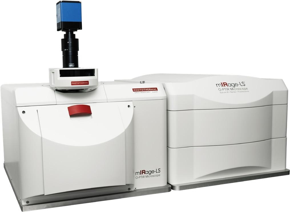
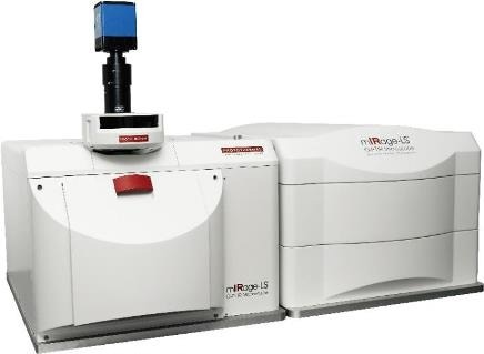
Figure 1.The mIRage-LS Sub-500nm IR multimodal microscope. Image Credit: Photothermal Spectroscopy Corp
With the recent introduction of O-PTIR spectroscopy with simultaneous Raman capabilities, conventional IR and Raman spectroscopy issues have been resolved. O-PTIR can now deliver submicrometer IR spatial resolution, without scattering artifacts, and is water-compatible with the ability to deliver IR and Raman spectroscopy simultaneously, providing the same spatial resolution from the same spot and the same time.
This makes it possible to utilize the real chemical complementarities of IR and Raman synergistically for the first time. These methods provide sub-cellular resolution combined with Raman and fluorescence imaging resolution, and can deliver broad macromolecular characterization on biologically relevant spatial scales (<500 nm).
For the first time, the incorporation of widefield epi-fluorescence imaging into this submicrometer simultaneous IR+Raman platform with the mIRage-LS product enables a sample registration free combination of these methods into a single instrument, heralding an advancement in life science research.
This novel product allows researchers to truly exploit these two techniques with potent synergy, accessing additional information and insights not available with either technique on its own while validating existing life science workflows.
包括新的反向传播模式,进一步改善了红外空间分辨率,开辟了新的O-PTIR光谱应用程序。
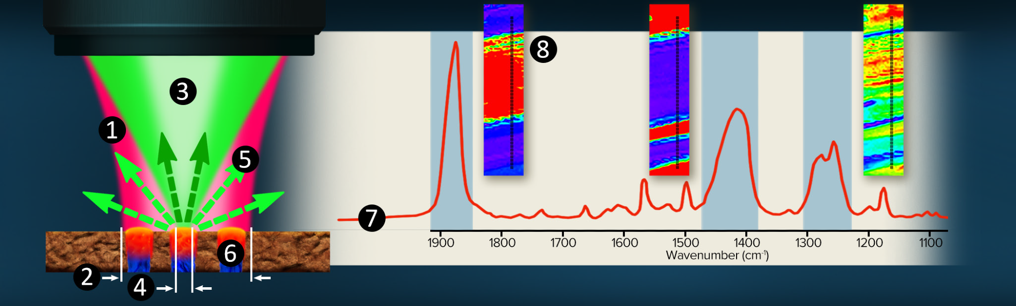
Figure 2.O-PTIR Co-propagating technique overview. Image Credit: Photothermal Spectroscopy Corp
打破IRSpatialResolutionBarrier with O-PTIR
With the advent of O-PTIR microspectroscopy, the 10-20 micrometer spatial resolution limitation of infrared spectroscopy has been removed, enabling IR to be used for sub-micrometer spatial resolution, high-resolution (<500nm) chemical structural imaging, and cellular and sub-cellular structures spectroscopy.
O-PTIR反射Mode共同PropagatingTechnique
The technique is illustrated in figure 2.
- A pulsed, tunable, infrared laser source (pump) is focused through a reflective (Cassegrain) objective.
- 红外光束聚焦为10-20 µm(衍射有限点)
- 共线性引入的可见激光器,通常是532 nm绿色拉曼激发激光(探针),通过相同的目标聚焦。
- The shorter wavelength green laser can be focussed to 30x tigher spot at around 500nm, allowing it to solely focuses on the blue region.
- As a function of IR laser wavelength adjustment, the green reflected (or transmitted) laser is detected.
- When the infrared wavelength overlaps an absorption band in the sample, energy is absorbed, resulting in a photothermal response and a change in the green reflectivity.
- Simultaneously monitoring the green laser reflectivity whilst tuning the IR source wavelenghts creates an infrared spectral response.
- 固定频率成像或高光谱数据集可用于映射化学功能组分布。
通过亚微米空间分辨率,通过检测反射(或传输)532 nm可见激光的情况,通过间接记录红外光谱。高质量(SNR)红外光谱可以在几秒钟内收集。
O-PTIRCounterPropagatingTechnique
Figure 3 illustrates the inclusion of a new high spatial resolution IR mode known as “counter-propagating”, another significant advancement with the mIRage-LS platform. Using regular high-power glass objectives as the probe delivery and collection optic is the key enabler of increased spatial resolution.
In contrast to the “co-propagating” mode, where the IR pump and visible probe beam are focused using all-reflective Cassegrain optics, the two beams are separated, with the IR beam being directed via the sample’s underside. This enables the use of high-power glass objectives for the delivery and collection of the probe beam.
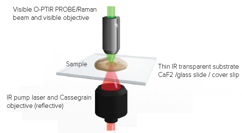
Figure 3.Counter propagating mode. IR light arrives from the bottom and the probe laser measures the sample response from the top with a high NA visible objective. Image Credit: Photothermal Spectroscopy Corp
The counter-propagating approach keeps all of the benefits of O-PTIR while significantly increasing spatial resolution and experimental versatility, with the ability to utilize water-dipping and oil-immersion objectives. Figure 4 depicts some of these novel forms of functioning.
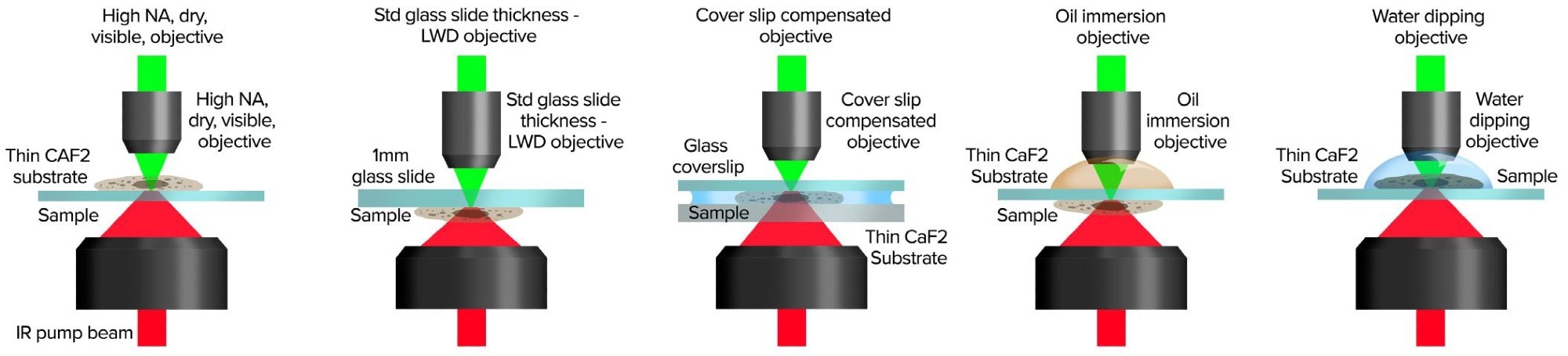
Figure 4.Counter-propagating mode provides application flexibility with a wide range of set ups for sample presentation. Image Credit: Photothermal Spectroscopy Corp
Simultaneous Raman/O-PTIRPrinciple ofOperation
The幻影-unique LS's idea and design, as shown in figure 5, allows Raman and O-PTIR to operate simultaneously on the same region on the sample, with identical spatial resolution.
研究人员可以使用两种方法的互补性质,并通过这些同时测量功能提供确认的光谱信息,以获得更精确的结果。
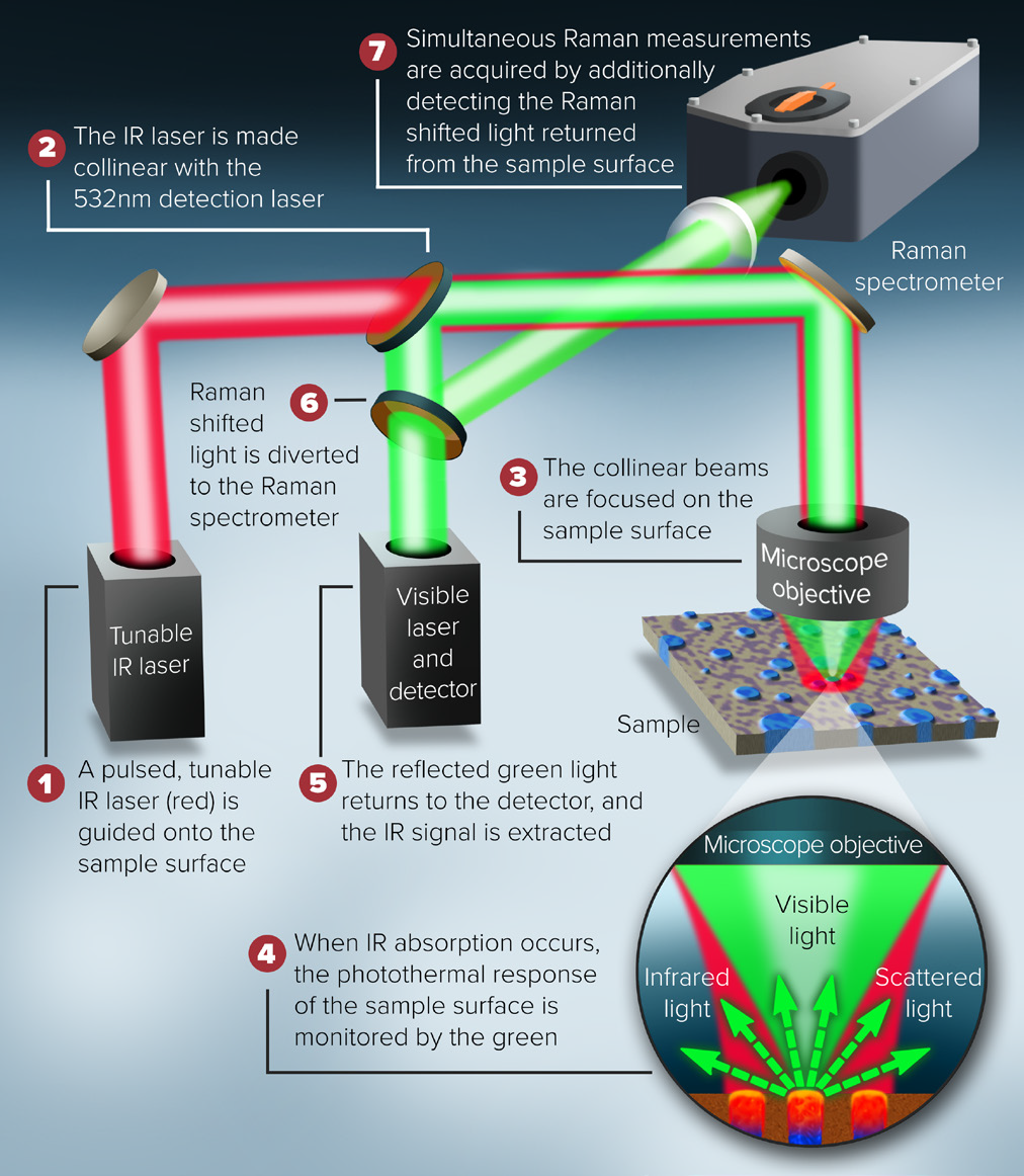
图5。O-PTIR和拉曼仪器组合的示意图。图片来源:光热光谱公司
幻影LSPrinciple ofOperation O-PTIR andFluorescence显微镜
Figure 6 depicts the system configuration for a combined O-PTIR sub-micron IR microscope, co-located with fluorescence microscopy. The pulsed IR beam is focused on the sample’s underside in counter-propagating mode. The fluorescence and visible probe beam paths are co-linear via the microscope objective enabling co-located measurements.
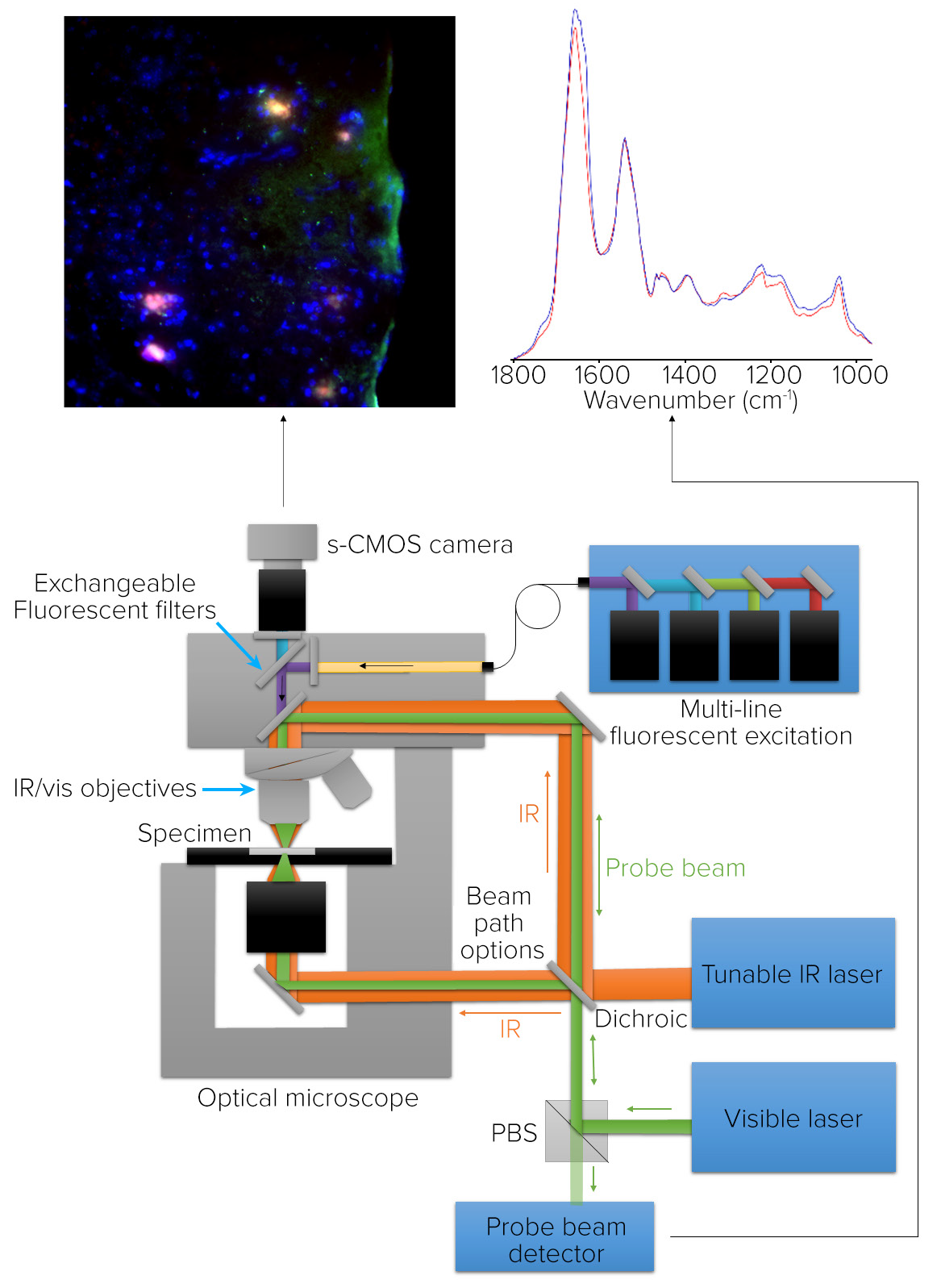
Figure 6.显微镜示意图,用于O-PTIR和共同定位的荧光显微镜。图片来源:光热光谱公司
O-PTIR uses high-resolution IR spectroscopy and imaging to quantify the same properties that the fluorescence microscope uses to identify biomolecules of interest with high molecular specificity.
To accommodate a variety of fluorescence applications, the fluorescence camera has an illumination component with a brightfield filter cube and a capacity for seven other filter cubes.
Cellular,蛋白质和Tissue申请with O-PTIR andCombinedTechniques
Neuroglioma Cells Characterization Using Counter-Propagating Mode
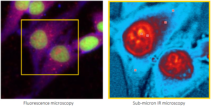
Figure 7 depicts the staining of neuroglioma cells with G3BP1 for protein stress granules, DAPI for nuclei, and BODIPY for lipids.
An RBG overlay widefield epifluorescence image with red displaying protein stress granules, blue revealing nucleus, and green indicating lipids is shown on the left. Square markers show O-PTIR spectral collecting sites. The brightfield image is located in the right.
The O-PTIR spectra, collected in a few seconds from the marker locations depicted in the left and right panes, are located at the bottom. It is possible to observe distinct spectrum changes compatible with the intended sub-cellular properties. The slight variation in the Amide I band of the protein stress granule, which indicates a distinct protein secondary structure from the other locations, is particularly noteworthy.
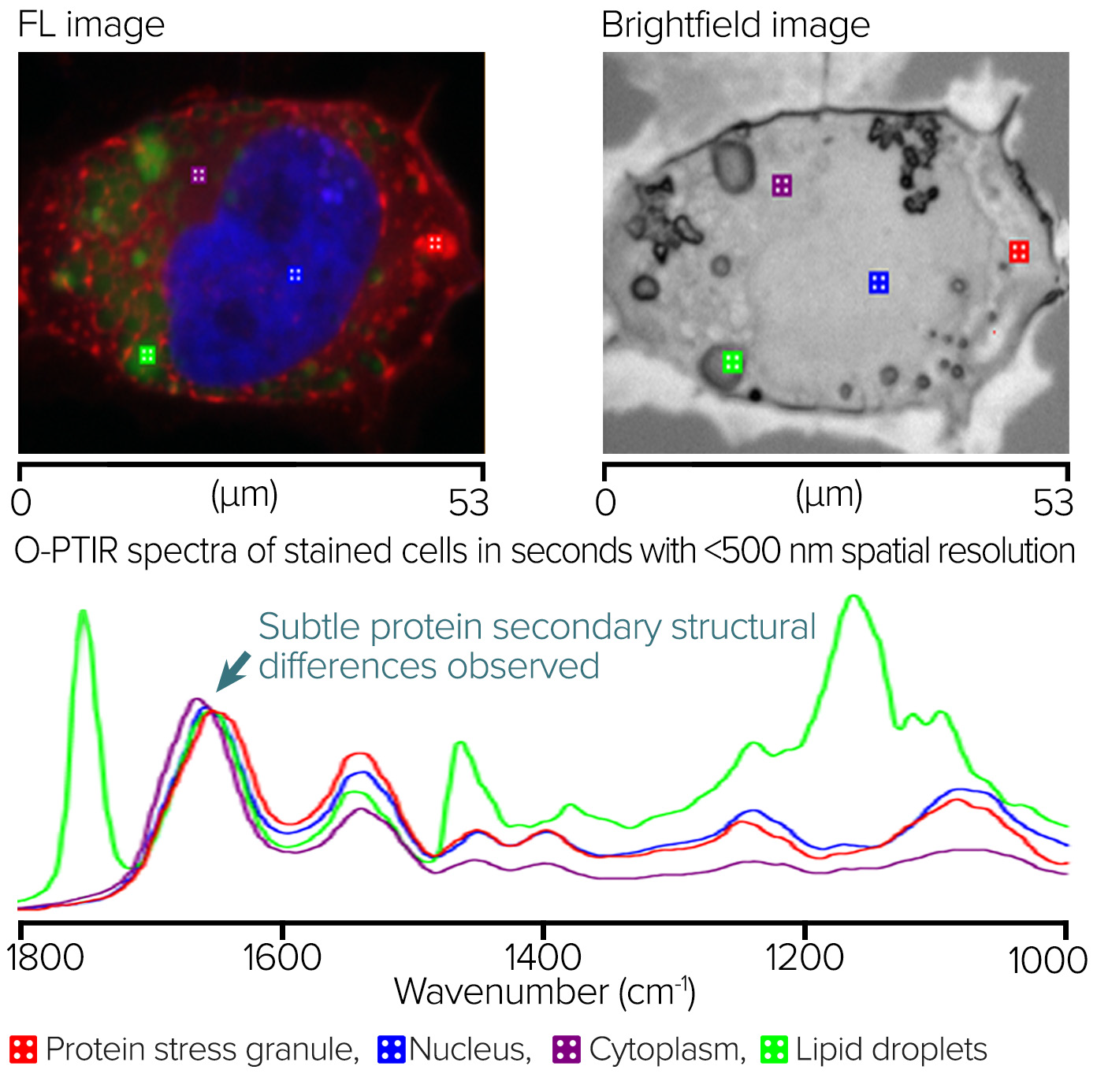
Figure 7.Fluorescence-guided Stained Neuroglioma cells and corresponding O-PTIR spectra. Image Credit: Photothermal Spectroscopy Corp
Blood Cells – Monolayer Measurement Using IR + Raman
Figure 8 depicts anIR + Ramanstudy of a monolayer of red blood cells collected in co-propagating mode from the surface of a glass microscope slide. The left pane image displays an optical image of a 70 µm x 70 µm region from which a Raman image was collected, with a false color displayed at 1583 cm-1using 532 nm excitation.
底部的底部O-PTIR和拉曼光谱是从选定的红细胞(亚微米分辨率)中获取的。该示例显着证明了IR和拉曼光谱法的互补性。血红蛋白蛋白带占主导地位的O-PTIR光谱,而血红素组的共振增强卟啉环振动主导了拉曼光谱。
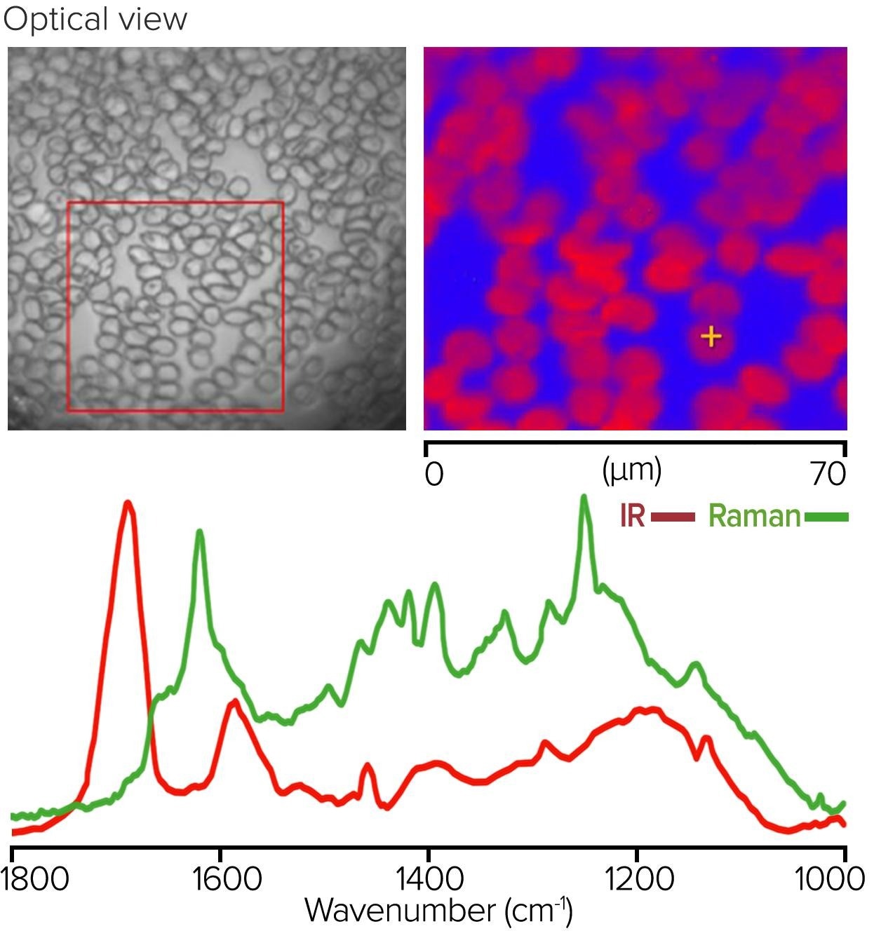
Figure 8.红细胞的单层,带有拉曼图像以及相应的O-PTIR和拉曼光谱。图片来源:光热光谱公司
Tissue Measurement with O-PTIR and Co-Located Fluorescence Microscopy
In a mouse model of Alzheimer's disease brain tissue slice, Amytracker 630 was used to identify the amyloid aggregates, AF488 was used to highlight proteins, and DAPI was used to identify the nucleus.
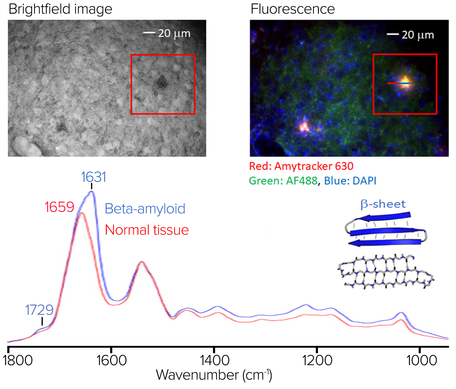
Figure 9.Stained tissue sample with fluorescence image and spectra showing significant spectral differences at 1631 cm-1. Image Credit: Photothermal Spectroscopy Corp
The top left of figure 9 depicts a brightfield image of the stained sample.
Figure 9 displays the RBG composite fluorescence image, which reveals amyloid aggregation in red/orange regions. Some amyloid aggregates identified in the fluorescence image are challenging to discern in the brightfield image.
The O-PTIR spectra averaged on (in blue) and off (in red) the aggregate are shown in figure 9, from the line profile indicated in the right pane. In the amide I band, where protein beta-sheet structures are characteristic, the aggregate's average spectrum exhibits various spectral variances and a prominent spectral signature at 1631 cm-1.
这体现了结合亚微米o-PTIR光谱的价值,然后在这种情况下,可以提供分子组成信息,在这种情况下,对蛋白质二级结构特别敏感,IR光谱与荧光成像的特征强度以突出显示淀粉样蛋白聚集区域在Brightfield显微镜中无法轻易看到其中。
盐水藻类
Both brightfield and autofluorescence (Ex 650 nm, Em 690 nm) images were captured using a 50 x, 0.8 NA objective in epi-fluorescence mode. Figure 10 illustrates how autofluorescence can display a wealth of contrast correlating to known sub-cellular structures, while the brightfield image is relatively feature-free.
Owing to its better contrast than the brightfield image, this autofluorescence image was used to direct the subsequent submicrometer IR and simultaneous Raman observations.
Despite the Raman signals being entirely overwhelmed by the sample’s autofluorescence, high-quality IR spectra devoid of any dispersive scattering artifacts (typical of direct IR methods such as FT-IR and QCL microscopy) could be produced in seconds from ~500 nm spots on the samples.
A simple lipid-to-protein ratio image was acquired at 1743 cm-1and 1655 cm-1using single frequency imaging mode for the lipid and protein to further investigate the macromolecular distributions.
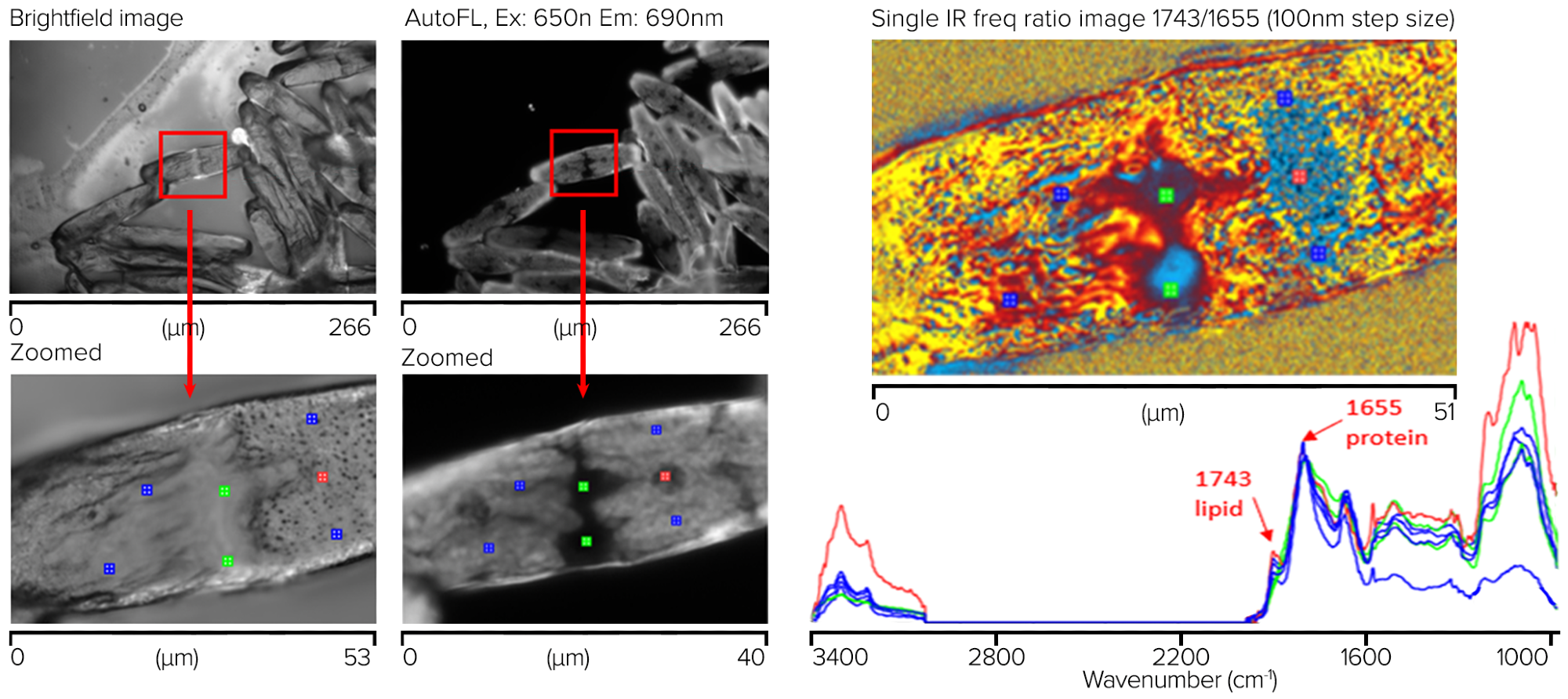
Figure 10.Saltwater algae sample showing measurement locations for corresponding spectra with chemical ratio image (1743/1655 cm-1). Image Credit: Photothermal Spectroscopy Corp
These results demonstrate the potential of this technique to investigate such samples for against external perturbations, such as nutrient depletion, changes in nutrients, and exposure to chemicals and drugs.
Single Bacterial Cell Using Simultaneous O-PTIR and Raman Spectroscopy
A single bacterial cell’s simultaneous O-PTIR and Raman spectra were acquired off a CaF2substrate. On the top left of figure 11, there is a visible image of bacterial cells. The orange box shows where the single-wavenumber IR image at 1655 cm-1并采用了50 nm的步长,突出了蛋白质成分的吸收。
底部图像显示同时从单个细菌细胞上的单个位置获取的同时量子计O-PTIR和拉曼光谱。使用覆盖3000-2700和1800-950 cm的双范围QCL在单个单元中获取O-PTIR光谱-1regions. The O-PTIR spectra were not post-processed, while the Raman spectra were baseline corrected.
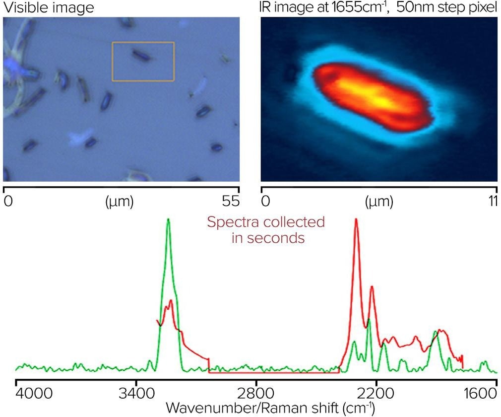
Figure 11.Single bacterial cell, with IR image and corresponding spectra from O-PTIR and Raman. Image Credit: Photothermal Spectroscopy Corp
FixedCellMeasurement withOilImmersionObjective
An oil-immersion objective (100x, 1.3 NA) was utilized in counter-propagating mode to measure IR spectral and images of a human cheek cell on an inverted glass coverslip to demonstrate the ultimate in optical and IR spatial resolution. Figure 12 illustrates how IR features as small as 285 nm can be observed in a rapid ultra-high-resolution image obtained by gathering molecularly specific single-frequency images, in this case at 1740 cm-1for lipid.
Even though the sample was mounted on glass, spectra are not entirely obscured by glass, unlike conventional IR methods such as FTIR and QCL microscopy. Instead, a wide silicate feature from the glass is observed, overlaid by cellular nucleic acid and carbohydrate characteristics. At the same time, the protein and lipid regions remain entirely free of any glass contributions.
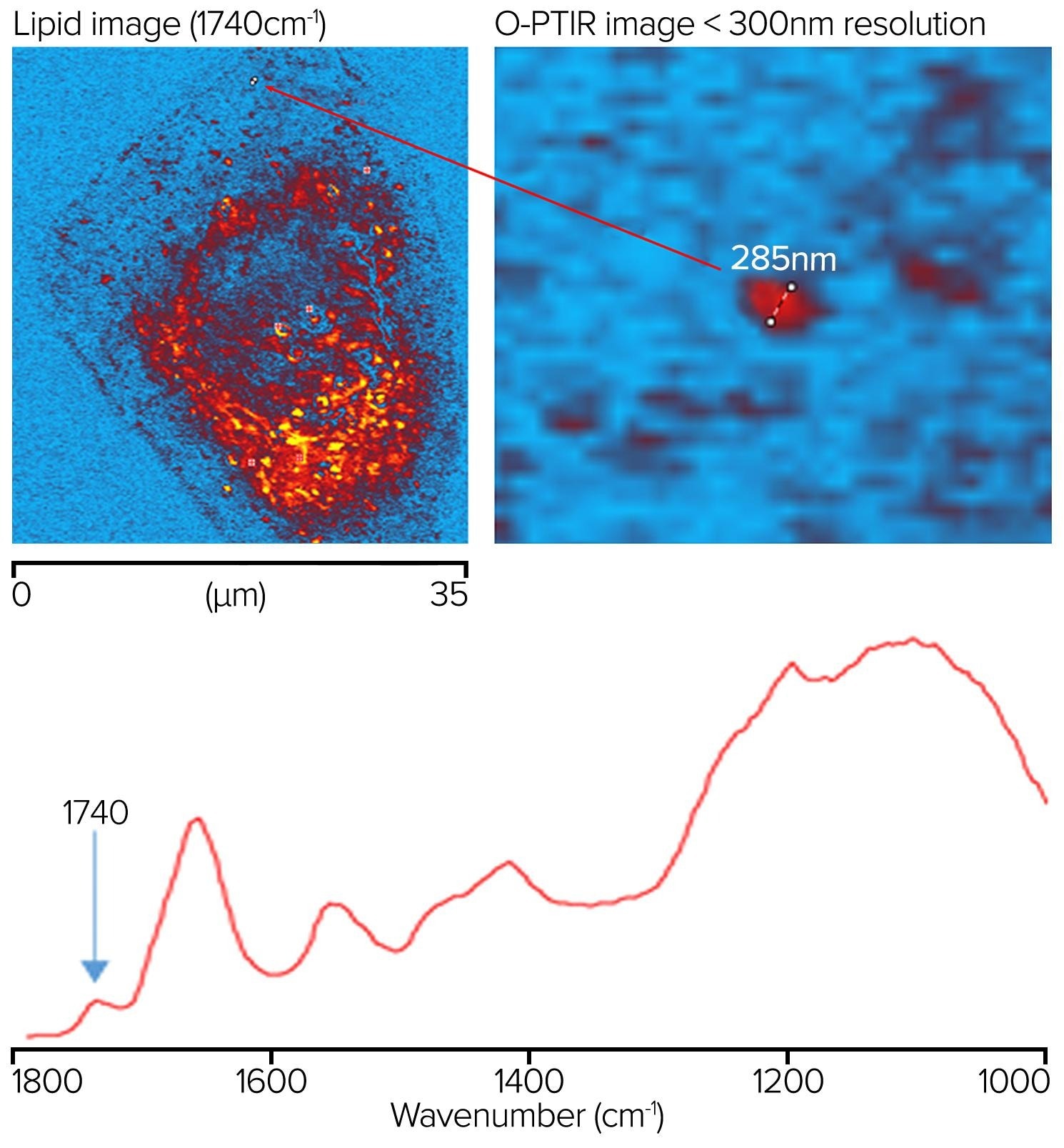
Figure 12.Lipid sample measured at 1740 cm-1showing a feature resolution of <300 nm with O-PTIR imaging. Image Credit: Photothermal Spectroscopy Corp
居住Cells inWater with O-PTIR andWater浸渍Objective
在300 µm厚的CAF上产生脸颊细胞2substrate. A drop of water was placed on the sample, and a reflecting Cassegrainian objective was used from beneath to deliver the IR light, with a visible water dipping objective being used to deliver and collected the probe beam in counterpropagating mode (refer back to the right side of figure 4). A clear image of the sample is displayed on the top left of figure 13.
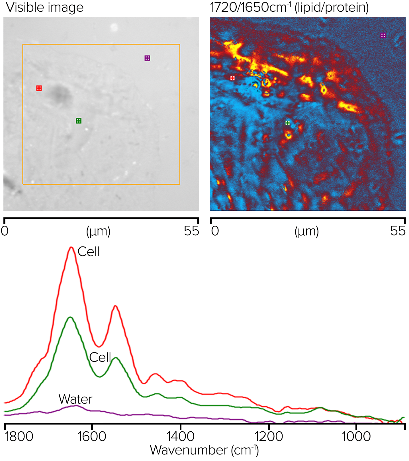
Figure 13.O-PTIR比率图像显示了相应的脂质/蛋白质区域和在著名位置进行测量的光谱。图片来源:光热光谱公司
The orange box indicates the region where single-wavenumber O-PTIR images were acquired with 50 nm spacing at 1720 and 1650 cm-1. The top right corner shows a ratio of these two images. The ratio image's red/orange parts reflect areas with greater lipid concentrations, whereas the blue sections have higher protein concentrations.
底部图显示了单点完整的O-PTIR光谱和适当颜色的光谱的位置。尽管没有使用水背景调节,但光谱几乎没有吸水。
Conclusion
This article demonstrates different life science applications ofO-PTIR microspectroscopy和与同时拉曼和荧光测量的成像。
The mIRage-LS product incorporates the simultaneous measurement of submicrometer O-PTIR and Raman spectra with widefield epi-fluorescence imaging, heralding a breakthrough for life science research by enabling the sample-registration-free combination of these techniques into a single instrument.
这一突破使研究人员能够通过强大的协同作用利用这两种技术,在与现有的生活科学工作流程对齐的同时,可以单独使用任何一种技术获得其他信息和见解。欧洲杯线上买球
幻影Product Overview
幻影®—亚微米红外光谱。图片来源:光热光谱公司
幻影®-R—Simultaneous sub-micron IR and Raman spectroscopy. Image Credit: Photothermal Spectroscopy Corp
幻影®-LS—New <500 nm IR with simultaneous Raman and co-located Fluorescence. Image Credit: Photothermal Spectroscopy Corp

该信息已从光热光谱公司提供的材料中采购,审查和调整。欧洲杯足球竞彩
有关此消息来源的更多信息,请访问Photothermal Spectroscopy Corp.